

Dysautonomia and the Upper Cervical Spine: What Your Neck May Have to Do With Your Autonomic Nervous System

If you've been diagnosed with dysautonomia — or if you've spent months or even years dealing with unexplained dizziness, heart racing, brain fog, and fatigue — you already know how frustrating this condition can be. What you may not know is that the answer to why your autonomic nervous system is misfiring could be sitting at the very top of your neck.
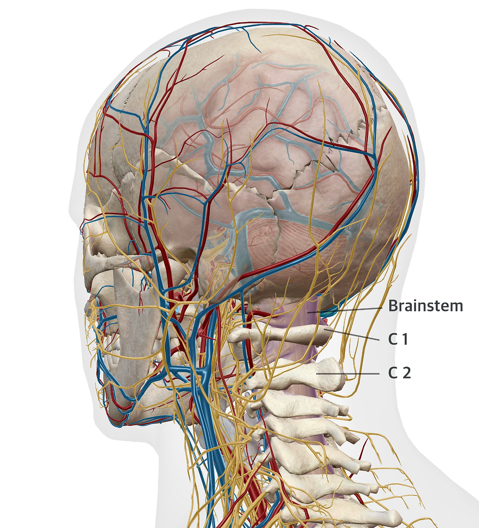
The craniocervical junction — where your skull meets the atlas (C1) and axis (C2) vertebrae — is one of the most neurologically significant regions in the entire body. It houses the brainstem, the vagus nerve, the vertebral arteries, and the sympathetic chain. When this area is misaligned, the consequences can ripple through every system your autonomic nervous system controls.
In this post, we'll explore what dysautonomia is, how it affects daily life, and why the connection between upper cervical misalignment and dysautonomia is gaining attention in both the medical and chiropractic communities.
What Is Dysautonomia?
Dysautonomia is an umbrella term for any condition in which the autonomic nervous system (ANS) doesn't work properly. Your ANS is essentially your body's autopilot — it controls heart rate, blood pressure, digestion, breathing, temperature regulation, bladder function, and even pupil dilation. When it malfunctions, patients can experience a wide and often bewildering range of symptoms that seem unrelated on the surface but share a common root: autonomic dysfunction.
There are several types of dysautonomia. The most common is Postural Orthostatic Tachycardia Syndrome (POTS), which causes the heart rate to spike by 30 or more beats per minute within 10 minutes of standing. Other forms include neurocardiogenic syncope (the most common cause of fainting), multiple system atrophy, pure autonomic failure, and autoimmune autonomic ganglionopathy. More recently, post-COVID dysautonomia has emerged as a significant concern, with studies showing that up to 30% of patients with severe long COVID develop POTS-like symptoms.

How Common Is Dysautonomia and How Serious Can It Be?
Dysautonomia affects over 70 million people worldwide, according to Dysautonomia International. In the United States alone, an estimated 1 to 3 million people live with POTS — making it more than twice as common as multiple sclerosis. Despite these numbers, the average patient waits 4 to 7 years before receiving a correct diagnosis. A 2025 study found the mean diagnostic delay was 7.7 years, and roughly 77% of POTS patients reported being told their symptoms were psychological before finally being diagnosed.
The severity of dysautonomia varies. Some patients experience mild, occasional symptoms. Others are profoundly disabled. Research published in the Journal of Internal Medicine found that POTS patients scored worse on quality-of-life measures than patients with diabetes, cardiovascular disease, COPD, HIV, or even most forms of cancer. Approximately 25% of POTS patients are unable to work, and over 70% report significant income loss due to their symptoms.
Symptoms can include heart palpitations, dizziness and lightheadedness, chronic fatigue, exercise intolerance, brain fog, nausea, temperature dysregulation, blurred vision, headaches, and gastrointestinal dysfunction. These symptoms don't just cause physical suffering — they can erode relationships, careers, and mental health when patients feel dismissed by doctors or unable to participate in normal daily activities.
Dysautonomia and the Upper Cervical Spine:
The Craniocervical Junction: A Neurological Crossroads
To understand why upper cervical misalignment matters for dysautonomia, you need to understand what lives at the top of your neck.
The craniocervical junction (CCJ) is formed by the base of the skull (the occiput), the atlas vertebra (C1), and the axis vertebra (C2). This small region is remarkable for several reasons. It allows roughly 40% of all neck flexion and extension and about 60% of all head rotation. But unlike the rest of the spine, there are no interlocking bones or intervertebral discs providing stability at C1-C2. Instead, the entire structure is held together by ligaments and muscles, making it inherently vulnerable to misalignment from trauma, repetitive stress, or connective tissue disorders.

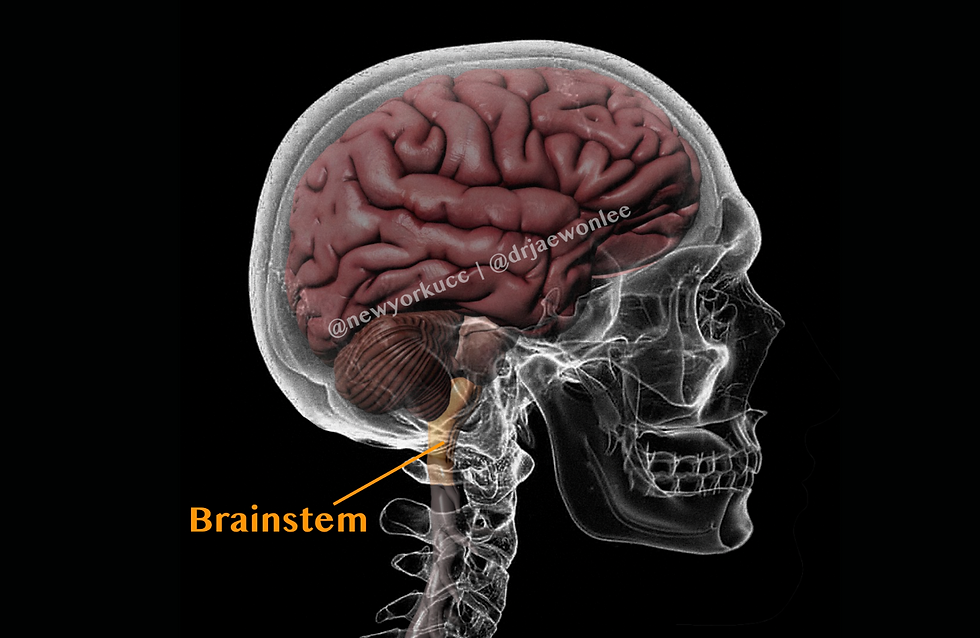
What makes this region critical for autonomic function is what it contains and what passes through it. The brainstem — specifically the medulla oblongata — transitions into the spinal cord right at this level. The medulla houses the body's primary autonomic control centers, including the cardiovascular center that regulates heart rate and blood pressure, the respiratory center, and the dorsal vagal complex. Any misalignment at C0-C2 that creates pressure, irritation, or altered blood flow at this level has the potential to directly affect how these control centers function.
How Upper Cervical Misalignment Can Disrupt Autonomic Function
There are several anatomical and neurological mechanisms through which a misalignment at the atlas or axis can contribute to dysautonomia symptoms.
The Vagus Nerve Connection
The vagus nerve — your body's longest cranial nerve and the primary parasympathetic pathway — originates in the medulla and exits the skull through the jugular foramen at the base of the skull. Its inferior ganglion (the nodose ganglion) sits directly in front of the atlas vertebra, separated by only millimeters of tissue. This means the vagus nerve is in intimate anatomical contact with C1. When the atlas shifts out of its proper position, it can mechanically compress or stretch the vagus nerve, reducing parasympathetic tone. The clinical result? Elevated heart rate, impaired digestion, reduced heart rate variability, and a cascade of autonomic symptoms that look very much like dysautonomia.

Brainstem Compression and Irritation
Even a subtle misalignment at C1-C2 can create mechanical deformation of the medulla and upper cervical cord where autonomic processing centers reside. This doesn't require a dramatic injury. The brainstem is remarkably sensitive to even low-grade, sustained pressure. Research by Milhorat et al. (2007) demonstrated that patients with connective tissue disorders showed occipitoatlantoaxial hypermobility with cranial settling — essentially, the skull sinking onto the spine — leading to brainstem compression and a significantly higher incidence of lower brainstem symptoms.
Vertebral Artery Compromise
The vertebral arteries take a uniquely winding path through the transverse foramina of the cervical vertebrae, making a particularly sharp turn around the lateral mass of the atlas before entering the skull. These arteries supply the brainstem and cerebellum through the vertebrobasilar system — in other words, they provide the blood supply to your autonomic control centers. Atlas misalignment can kink, compress, or create torsion in these arteries, potentially reducing blood flow to the very structures that regulate your heart rate and blood pressure.

CSF Flow Disruption
The craniocervical junction is also a critical "bottleneck" for cerebrospinal fluid (CSF) flow between the brain and the spinal canal. Research by Flanagan (2015) and Rosa et al. (2018) has shown that misalignment at C0-C1 or C1-C2 can obstruct normal CSF flow through the foramen magnum. This disruption may contribute to intracranial pressure changes, impaired waste clearance from the brain, and neurological symptoms including brain fog and headaches — symptoms that are extremely common in dysautonomia.

Sympathetic Nervous System Overactivation
The superior cervical sympathetic ganglion — the highest ganglion in the sympathetic chain — sits directly in front of the C2-C3 vertebral bodies. This ganglion communicates with the vagus nerve, meaning both branches of the autonomic nervous system converge at the C1-C2 region. Chronic upper cervical misalignment may create sustained sympathetic overdrive by irritating this ganglion, reducing vagal tone through mechanical interference, and disrupting the descending autonomic pathways at the brainstem level.
What Does the Research Say About Upper Cervical Chiropractic and Dysautonomia?
The most significant study in this area is the landmark Bakris et al. (2007) study, published in the Journal of Human Hypertension (a Nature Publishing Group journal). This double-blind, placebo-controlled pilot study randomized 50 patients with Stage 1 hypertension to either atlas adjustment or a sham procedure. After 8 weeks, the group receiving the real atlas correction showed systolic blood pressure reductions of 17 mmHg — equivalent to taking two blood pressure medications simultaneously — with no adverse effects. The authors concluded that "restoration of Atlas alignment is associated with marked and sustained reductions in BP."
Studies examining heart rate variability (HRV) — a direct measure of autonomic nervous system balance — have also shown promising results. Welch and Boone (2008) demonstrated that cervical adjustments produced a shift toward parasympathetic dominance, while a randomized crossover study by Win et al. (2015) found that upper cervical manipulation at C1-C2 specifically enhanced parasympathetic function with significant blood pressure decreases.
A 2022 case study published in the Journal of Family Medicine and Primary Care documented a 50-year-old woman with three years of POTS symptoms who achieved complete resolution of her dizziness, neck pain, and POTS after three months of chiropractic rehabilitation, with results maintained at 12-month follow-up.
It's important to be transparent: while the biological plausibility for upper cervical effects on autonomic function is strong, large-scale randomized controlled trials specifically targeting POTS and dysautonomia have not yet been published. The existing evidence — from controlled blood pressure studies, HRV research, case reports, and anatomical science — is promising and growing, but more research is needed. Upper cervical care is best understood as a complementary approach that addresses a potential structural contributor to autonomic dysfunction, not a standalone cure.
Unusual Connections You Might Not Expect
The Ehlers-Danlos Syndrome–POTS–Craniocervical Instability Triad
One of the most fascinating emerging connections involves Ehlers-Danlos Syndrome (EDS), a group of connective tissue disorders that cause ligamentous laxity throughout the body. When the ligaments at the craniocervical junction become too loose, the result is craniocervical instability (CCI) — and a surprisingly high rate of dysautonomia. Dr. Fraser Henderson's 2021 study in World Neurosurgery found that among 20 EDS patients with atlantoaxial instability, every single patient had lightheadedness, and 15 had refractory syncope despite maximal medical management. After surgical stabilization of C1-C2, patients showed statistically significant improvement. This suggests that some cases of "unexplained" POTS may actually have a structural origin at the craniocervical junction.
Coat Hanger Pain: A Diagnostic Clue in Disguise
Many dysautonomia patients experience a distinctive aching pain that spans the neck, shoulders, and upper back in the shape of a coat hanger. This "coat hanger pain" affects up to 93% of patients with pure autonomic failure and is caused by inadequate blood flow to the muscles that constantly work to support the head. Unlike typical mechanical neck pain, coat hanger pain reliably improves when lying down and worsens when standing. It's frequently misdiagnosed as fibromyalgia or tension headaches, but recognizing it can be an important clue pointing toward autonomic dysfunction.
Why a 2mm Shift at C1 Can Affect Your Whole Body
The atlas vertebra weighs only about 2 ounces, but it supports a skull weighing 10 to 12 pounds. With no intervertebral disc at C1-C2, structural stability depends entirely on ligaments. The vagus nerve's nodose ganglion and the sympathetic chain's superior cervical ganglion both sit millimeters from these vertebrae. The Bakris study showed that correcting atlas lateral displacement from just over one degree to near-zero produced cardiovascular changes rivaling pharmaceutical intervention. At the craniocervical junction, even very small structural shifts can have outsized neurological consequences.
How Upper Cervical Chiropractic Care Differs From Traditional Chiropractic
Upper cervical chiropractic is a specialized discipline that focuses exclusively on the relationship between the skull, atlas, and axis. It differs from general chiropractic in several important ways.
First, the adjustments are precise and specifically targeted to the craniocervical junction. Second, every correction is calculated from advanced imaging specific to the individual patient's anatomy. No two atlas misalignments are the same, and the correction vector is mathematically determined for each person. Third, patients are only adjusted when objective testing — including postural analysis and thermographic scanning — confirms that a misalignment is present. The goal is for corrections to hold over time; needing fewer adjustments is a sign of progress, not a reason for more visits.
For dysautonomia patients, this gentle, precision-based approach is particularly important. Many patients with autonomic dysfunction also have heightened sensitivity, and the low-force nature of upper cervical techniques is generally well-tolerated even by those who have struggled with other forms of treatment.
Dysautonomia Treatment in New York: Finding the Right Support
If you're living with dysautonomia, POTS, or unexplained autonomic symptoms in New York, you deserve a care team that looks beyond medication management alone. While conventional treatments — including increased salt and fluid intake, compression garments, structured exercise programs, and medications like fludrocortisone, midodrine, or propranolol — remain important foundations of care, they don't address potential structural contributors to autonomic dysfunction.
At New York Upper Cervical Chiropractic, we specialize in precise, gentle corrections of the atlas and axis vertebrae using advanced imaging and objective neurological testing. Our office serves patients throughout New York City, Manhattan, Long Island, Queens, Brooklyn, Nassau County, Westchester, and the surrounding areas who are searching for dysautonomia treatment, POTS relief, autonomic dysfunction support, and upper cervical chiropractic care. Whether you've been recently diagnosed or have been struggling with symptoms for years, we're here to evaluate whether an upper cervical misalignment may be contributing to your condition and to work alongside your existing medical team to support your recovery.
If you or someone you love is dealing with dysautonomia, POTS, chronic dizziness, heart palpitations, brain fog, or other autonomic symptoms, we invite you to schedule a consultation. Sometimes the missing piece isn't another medication — it's the structural integrity of the most neurologically important junction in your body.
FAQs About Dysautonomia and Upper Cervical Chiropractic
1. Can upper cervical chiropractic cure dysautonomia?
Upper cervical chiropractic is not a cure for dysautonomia. However, if an upper cervical misalignment is contributing to brainstem irritation, vagus nerve interference, or disrupted blood flow to the autonomic control centers, correcting that misalignment may help reduce symptoms. It works best as a complementary approach alongside conventional medical treatment, not as a replacement for it.
2. How do I know if my atlas is misaligned?
Only specialized imaging and a thorough upper cervical examination can confirm atlas misalignment. Common indicators include a history of head or neck trauma (car accidents, falls, concussions, sports injuries), chronic headaches or migraines, uneven shoulders or hips, and symptoms that began or worsened after an injury. Many people with significant misalignment experience no neck pain at all.
3. Is upper cervical chiropractic safe for people with POTS or EDS?
Specific upper cervical techniques are generally well-tolerated by POTS patients. For patients with Ehlers-Danlos Syndrome, care should be provided by a practitioner experienced with hypermobility conditions. Patients with vascular EDS should consult their geneticist before pursuing any manual therapy. Aggressive rotational neck manipulation is not used in upper cervical chiropractic and is not appropriate for these patients.
4. How long does it take to see results?
Results vary from patient to patient. Some people notice improvement within the first few visits, while others require several weeks or months of care. An initial phase of more frequent visits typically transitions into a stabilization period and eventually maintenance care. The objective is for corrections to hold longer over time, which means fewer visits as your body stabilizes.
5. Can a neck injury cause dysautonomia?
Yes, there is growing evidence that head and neck injuries — including whiplash, concussions, and falls — can trigger autonomic dysfunction. A 2018 systematic review found that 33 of 36 studies identified autonomic abnormalities in concussed populations, and data from a large pediatric institution showed that over 11% of POTS diagnoses had symptom onset within three months of a concussion. The mechanism likely involves injury to both the brainstem and the cervical structures that house the vagus nerve and sympathetic chain.
New York Upper Cervical Chiropractic
📍 505 Northern Blvd, Suite 309, Great Neck, NY 11021
📞 516) 969-3330
@ drjaewonlee | @newyorkucc
References
Bakris G, Dickholtz M Sr, Meyer PM, et al. Atlas vertebra realignment and achievement of arterial pressure goal in hypertensive patients: a pilot study. Journal of Human Hypertension. 2007;21(5):347-352.
Shaw BH, Stiles LE, Bourne K, et al. The face of postural tachycardia syndrome — insights from a large cross-sectional online community-based survey. Journal of Internal Medicine. 2019;286(4):438-448.
Bourne KM, Stiles LE, Shaw BH, et al. Postural orthostatic tachycardia syndrome is associated with significant employment and economic loss. Journal of Internal Medicine. 2021;290(1):203-212.
Milhorat TH, Bolognese PA, Nishikawa M, et al. Syndrome of occipitoatlantoaxial hypermobility, cranial settling, and Chiari malformation type I in patients with hereditary disorders of connective tissue. Journal of Neurosurgery: Spine. 2007;7(6):601-609.
Henderson FC, et al. Refractory syncope and presyncope associated with atlantoaxial instability: preliminary evidence of improvement following surgical stabilization. World Neurosurgery. 2021;149:e854-e865.
Welch A, Boone R. Sympathetic and parasympathetic responses to specific diversified adjustments to chiropractic vertebral subluxations of the cervical and thoracic spine. Journal of Chiropractic Medicine. 2008;7(3):86-93.
Win N, Jorgensen A, Chen Y, Haneline M. Effects of upper and lower cervical spinal manipulative therapy on blood pressure and heart rate variability. Journal of Chiropractic Medicine. 2015;14(1):1-9.
Flanagan MF. The role of the craniocervical junction in craniospinal hydrodynamics and neurodegenerative conditions. Neurology Research International. 2015;2015:794829.
Rosa S, Baird JW, Harshfield D, Chehrenama M. Craniocervical junction syndrome: anatomy of the craniocervical and atlantoaxial junctions and the effect of misalignment on cerebrospinal fluid flow. IntechOpen. 2018.
Chu ECP, Lin AFC. Relief of postural orthostatic tachycardia syndrome with chiropractic rehabilitation. Journal of Family Medicine and Primary Care. 2022;11(7):4006-4009.
Kingston et al. Effectiveness of spinal manipulation in influencing the autonomic nervous system — a systematic review and meta-analysis. Journal of Manual & Manipulative Therapy. 2024;32(1).
Woodfield HC, et al. Craniocervical chiropractic procedures — a précis of upper cervical chiropractic. Journal of the Canadian Chiropractic Association. 2015;59(2):173-192.
Disclaimer: This blog post is for educational purposes only and is not intended as medical advice. Dysautonomia is a complex condition that should be evaluated by a qualified healthcare provider. Upper cervical care may be considered as part of a comprehensive approach. Please consult with your healthcare provider to determine what approach is best for you.


